
Circulatory Heart Diagram
Follow a drop of blood through the heart. The interactive diagram labels the four chambers, the valves and the major blood vessels; step through the circulation to see blood travel from the body to the lungs and back, and switch to quiz mode to test yourself. It is a simplified teaching schematic — perfect for biology classes, revision and nursing prerequisites.
- Free to use
- No sign-up
- Runs in your browser
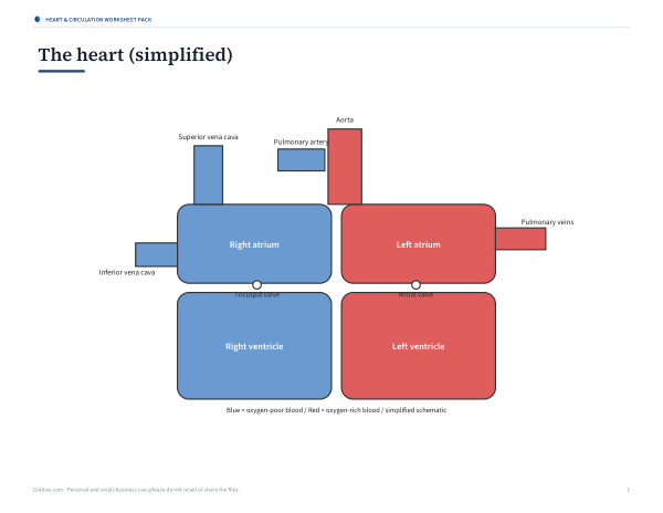
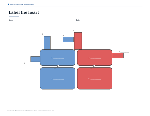
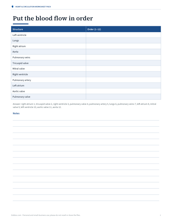
Heart and blood flow diagram
Heart & Circulation Worksheet Pack
Printable labelled heart diagram, a blank labelling worksheet with answers, a blood flow sequencing activity, a vessels comparison chart and a revision quiz.
- Labelled heart (PDF)
- Blank worksheet (PDF)
- Sequencing (PDF/DOCX)
- Vessels chart (PDF)
- Revision quiz (PDF, DOCX)
Formats: PDF, DOCX. Instant download after payment (link valid 72 hours, up to 5 downloads). AI-assisted: the templates were drafted with AI help and reviewed and laid out before publishing.
$3.00 USD, one-time 5 files · about $0.60 each
- Instant download
- 7-day refund
- Secure checkout by Stripe
7-day money-back guarantee. Full refund within 7 days of purchase, even after you download.
Free sample: Blood Flow Sequencing (PDF, 54 KB)
Full refund within 7 days — see the refund policy and license.
Free printables
Single sheets you can print right now, no sign-up. The full pack adds every sheet in one download plus the editable files.
 Printable Vessels Chart
 Printable Circulation Revision Quiz
The heart at a glance
The heart is a muscular pump about the size of a fist, sitting slightly left of centre in the chest. It has four chambers: two upper atria that receive blood and two lower ventricles that pump it out. The right side handles oxygen-poor blood and sends it to the lungs; the left side handles oxygen-rich blood and sends it to the whole body. Four valves keep blood moving in one direction. At rest, an adult heart typically beats about 60–100 times a minute.
Blood flow through the heart, step by step
- Oxygen-poor blood from the body enters the right atrium through the superior and inferior vena cava.
- It passes through the tricuspid valve into the right ventricle.
- The right ventricle pumps it through the pulmonary valve into the pulmonary artery.
- In the lungs, blood releases carbon dioxide and picks up oxygen.
- Oxygen-rich blood returns through the pulmonary veins to the left atrium.
- It passes through the mitral (bicuspid) valve into the left ventricle.
- The left ventricle pumps it through the aortic valve into the aorta.
- Arteries carry it to the body’s organs and tissues; veins bring it back to the heart.
Two circuits: pulmonary and systemic
| Circuit | From | To | Purpose |
|---|---|---|---|
| Pulmonary | Right ventricle | Lungs → left atrium | Exchange carbon dioxide for oxygen |
| Systemic | Left ventricle | Body → right atrium | Deliver oxygen and nutrients, collect waste |
Because blood passes through the heart twice on each complete circuit, humans have a double circulatory system. The left ventricle has a much thicker wall than the right because it must push blood around the whole body.
Arteries, veins and capillaries
| Vessel | Carries blood | Features |
|---|---|---|
| Arteries | Away from the heart | Thick, elastic, muscular walls; high pressure |
| Veins | Towards the heart | Thinner walls, valves to prevent backflow; low pressure |
| Capillaries | Between arteries and veins | One cell thick; where exchange with tissues happens |
The pulmonary artery and pulmonary veins are the exceptions to the “arteries carry oxygen-rich blood” rule: the pulmonary artery carries oxygen-poor blood to the lungs, and the pulmonary veins carry oxygen-rich blood back.
Why diagrams show left and right reversed
Heart diagrams are drawn as if you are facing the person, so the heart’s right side appears on the left of the page. That is why the right atrium and right ventricle are on the left in the interactive diagram and in most textbooks.
The heartbeat and valves
Each heartbeat has two main phases: systole, when the ventricles contract and push blood out, and diastole, when they relax and fill. The “lub-dub” sound comes from valves closing: “lub” as the tricuspid and mitral valves close at the start of ventricular contraction, and “dub” as the pulmonary and aortic valves close when it ends. The heart’s own electrical system, starting at the sinoatrial node, keeps the beat coordinated.
Worked example: tracing one circuit
Start at the right atrium and press “Next step”. The yellow marker moves through the tricuspid valve to the right ventricle, out through the pulmonary valve and artery to the lungs, back through the pulmonary veins to the left atrium, through the mitral valve into the left ventricle, then through the aortic valve into the aorta and on to the body, before returning through the vena cava. Saying each step aloud while the marker moves is an effective way to memorise the order.
Blood pressure and pulse
Each contraction of the left ventricle sends a pressure wave along the arteries, which you can feel as your pulse at the wrist or neck. Blood pressure is written as two numbers: the higher systolic pressure during contraction and the lower diastolic pressure during relaxation. Measuring your pulse — counting beats for 30 seconds and doubling — is a simple classroom experiment, and comparing resting and post-exercise pulse shows how the heart responds to the body’s needs.
Key vocabulary
- Atrium (plural atria) — upper receiving chamber.
- Ventricle — lower pumping chamber.
- Septum — wall dividing the left and right sides of the heart.
- Valve — flap that keeps blood flowing one way.
- Oxygenated / deoxygenated — blood rich or poor in oxygen.
- Coronary arteries — vessels supplying the heart muscle itself.
Keeping the heart healthy
- Regular physical activity strengthens the heart muscle.
- A balanced diet with plenty of fruit, vegetables and whole grains supports heart health.
- Not smoking is one of the most important steps for heart health.
- Managing blood pressure and cholesterol reduces strain on the heart.
- Sleep and stress management matter too.
This page is for education only. For symptoms such as chest pain, shortness of breath or palpitations, seek medical advice promptly.
Privacy
The diagram runs in your browser.
Frequently asked questions
What are the four chambers of the heart?
Right atrium, right ventricle, left atrium and left ventricle.
What is the order of blood flow through the heart?
Vena cava → right atrium → tricuspid valve → right ventricle → pulmonary valve → pulmonary artery → lungs → pulmonary veins → left atrium → mitral valve → left ventricle → aortic valve → aorta → body.
Why is the left ventricle wall thicker?
It pumps blood to the whole body at higher pressure.
Which artery carries oxygen-poor blood?
The pulmonary artery.
What makes the heartbeat sound?
The heart valves closing.
Why is the heart’s right side on the left of diagrams?
Diagrams show the heart as if you are facing the person.
How many times does the heart beat in a day?
Roughly 100,000 times, at about 70 beats per minute.
What supplies the heart muscle with blood?
The coronary arteries.
Is this medical advice?
No, it is an educational diagram.